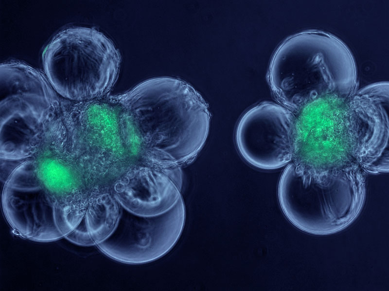
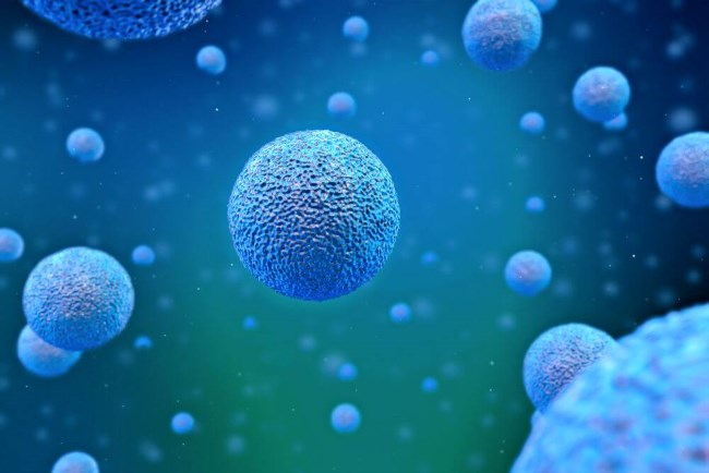
STEM CELLS
Stem Cells can only be ordered by an U.S. licensed physician with a valid NPI number. Product ordered and paid for will be shipped directly from the manufacturer's warehouse to the physicians practice. Once Product has left the warehouse it cannot be returned for a refund, as the chain of custody has been broken.
If you're looking for stem cell treatments, MEDICUS LLC is a strategic partner for physicians in your community that have decided to educate the public on cutting-edge regenerative cellular medicine. Our purpose is to help answer questions concerning Regenerative Cell Therapy to improve the lives of patients all over the United States. Patients can trust MEDICUS LLC to help them find physicians to help achieve their wellness goals and find the path to optimal wellness. In order to provide relief for a variety of issues, our partner physicians take an integrated approach to health care that treats patients like whole people rather than a collection of symptoms. For instance, for those suffering from chronic pain, our partner physicians work to address the source of your problem, rather than simply treating the symptoms. Because of this dedication to finding the root cause of your medical issues, with advanced Regenerative Cellular Medicine, chronic joint pain and other degenerative health problems are solved by utilizing regenerative cell treatments. There's no better place to find answers for conditions that include but are not limited to:
- Back Pain & Neck Pain
- Arthritis Pain
- Muscle & Ligament Injuries
- Shoulder Pain and Joint Injuries
- Knee Pain and Knee Arthritis
- Sciatica Leg Pain
- Peripheral Neuropathy
- Disc Herniations and Disc Bulges
- Spinal Stenosis
- Failed Back Surgery Treatment
At MEDICUS LLC, we provide regenerative medicine education and referral to qualified trained physicians that patients can trust. If you're seeking a solution for a medical problem like chronic pain, it's time to attend one of our educational seminars.
CALL US AT (855) go2-MEDI or (855) 462-6334 FOR MORE INFORMATION OR TO GET CONNECTED TO ONE OF OUR MEDICAL DOCTORS TO DISCUSS YOUR INDIVIDUAL NEEDS AND TREATMENT OPTIONS.
RESEARCH has been done on:
INFLAMMATION & FIBROSIS:
Amniotic membrane and amniotic cells: Potential therapeutic tools to combat tissue inflammation and fibrosis?
Placenta. 2011 Oct;32 Suppl 4:S320-5. doi: 10.1016/j.placenta.2011.04.010. Epub 2011 May 12. Manuelpillai U1, Moodley Y, Borlongan CV, Parolini O. 1Monash Institute of Medical Research, Monash University, Clayton, Victoria 3168, Australia.
Abstract
In addition to the placenta, umbilical cord and amniotic fluid, the amniotic membrane is emerging as an immensely valuable and easily accessible source of stem and progenitor cells. This concise review will focus on the stem/progenitor cell properties of human amniotic epithelial and mesenchymal stromal cells and evaluate the effects exerted by these cells and the amniotic membrane on tissue inflammation and fibrosis.Copyright Ⓒ 2011 Elsevier Ltd. All rights reserved. PMID: 21570115 [PubMed - indexed for MEDLINE]
Review preclinical studies on placenta-derived cells and amniotic membrane: An update
Placenta. 2011 Mar;32 Suppl 2:S186-95. doi: 10.1016/j.placenta.2010.12.016. Epub 2011 Jan 19. Parolini O1, Caruso M. 1Centro di Ricerca E. Menni, Fondazione Poliambulanza-Istituto Ospedaliero, Brescia, Italy.
Abstract
Recent years have seen considerable advances in our knowledge of the biology and properties of stem/progenitor cells isolated from placental tissues. This has encouraged researchers to address the potential effects of these cells in animal models of different diseases, resulting in increasing expectations regarding their possible utility for cell-based therapeutic applications. This rapidly evolving research field is also enriched by studies aimed at expanding the use of the whole amniotic membrane (AM), a well-known surgical material, for pathological conditions other than those tested so far and for which clinical applications already exist. In this review, we provide an update on studies that have been performed with placenta-derived cells and fragments of the entire AM to validate their potential clinical applications in a variety of diseases, in particular those associated with degenerative processes induced by inflammatory and fibrotic mechanisms. We also offer, as far as possible, insight into the interpretation and suggested mechanisms to explain the most important outcomes achieved to date.Copyright Ⓒ 2011 Elsevier Ltd. All rights reserved. PMID: 21251712 [PubMed - indexed for MEDLINE]
Suppression of inflammatory and fibrotic responses in allergic inflammation by the amniotic membrane stromal matrix
Clin Exp Allergy. 2005 Jul;35(7):941-8.Solomon A1, Wajngarten M, Alviano F, Anteby I, Elchalal U, Pe'er J, Levi-Schaffer F. 1Department of Ophthalmology, Hadassah University Hospital, The Hebrew University-Hadassah Faculty of Medicine, Jerusalem, Israel.
Abstract
BACKGROUND: The amniotic membrane (AM), which is the innermost layer of the placenta, was shown to possess anti-inflammatory and anti-fibrotic properties in various in vitro and clinical studies.PURPOSE: To evaluate the anti-fibrotic and anti-inflammatory effects of the AM matrix (AMM) on human conjunctival and lung fibroblasts in an in vitro system that tests fibrotic and inflammatory responses at the effector stages of allergic inflammation.
METHODS: Human conjunctival or lung fibroblasts were seeded on plastic or on the stromal aspect of the AM, which was mounted on plastic inserts. Sonicates of human peripheral blood eosinophils activated with lipopolysaccharide (LPS), or human mast cell (HMC-1) leukaemia cell sonicates, were added to sub-confluent fibroblast monolayers. Proliferation of the sub-confluent fibroblasts was assessed using the [3H]-thymidine incorporation assay. The production of transforming growth factor (TGF)-beta1, granulocyte-macrophage colony-stimulating factor (GM-CSF) and IL-8 in conjunctival or lung fibroblasts was measured in conditioned media from these cultures by ELISA.
RESULTS: After 4 days in culture, the [3H]-thymidine incorporation assay indicated a reduced proliferation of activated conjunctival and lung fibroblasts when cultured directly on the AMM. The production of both TGF-beta1 and IL-8 was significantly suppressed in activated conjunctival fibroblasts cultured on the AMM compared with those cultured on plastic, while the production of both TGF-beta1 and GM-CSF was decreased in human lung fibroblast cultured on the AMM.
CONCLUSIONS: The AMM is capable of suppressing fibrotic responses in an in vitro system of effector stages of ocular allergic inflammation. These data may provide a basis for exploring matrix components in the AM for the treatment of allergic eye disease.
PMID: 16008682 [PubMed - indexed for MEDLINE]
SPINE:
Implantation of amniotic membrane to reduce postlaminectomy epidural adhesions.
Eur Spine J. 2009 Aug;18(8):1202-12. doi: 10.1007/s00586-009-1013-x. Epub 2009 Apr 30.Tao H1, Fan H.
1Department of Orthopaedics and Traumatology, Xi-jing Hospital, The Fourth Military Medical University, 710032, Xi'an, China.
Abstract
Postlaminectomy epidural adhesion is implicated as a main cause of "failed back surgery syndrome" and associated with increased risk of complications during revision surgery. Various materials acting as mechanical barriers to reduce fibroblasts infiltration into epidural space have met with limited success. In present research, amniotic membrane (AM) was studied to investigate its effects on reducing epidural scar adhesion after laminectomy in a canine model. Laminectomy sites were created at L-1, L-3, L-5, and L-7 levels in 24 adult mongrel dogs. Freeze dried AM (FAM), cross-linked AM (CAM), and autologous free fat (AFF) were implanted, respectively, at a randomly assigned site in each dog with the remaining untreated site serving as internal control. The animals were sacrificed at 1, 6, and 12 weeks postoperatively. Then, gross pathologic observation including scar amount and adhesion tenacity, qualitative histology evaluation, and quantitative histology analysis were compared. Gross observation demonstrated that scar amount and adhesion tenacity of CAM group were significantly lower in comparison with those of FAM and non-treatment groups. A white, slightly vascularized CAM layer covered the dura mater without tenacious scar adhesion. The histology analysis also indicated reduced fibroblasts infiltration and consequent epidural fibrosis, which were similar to the results of AFF group. In conclusion, the CAM is effective in reducing epidural fibrosis and scar adhesion after laminectomy in canine model. It is a promising biomaterial for future clinical applications.PMID: 19404691 [PubMed - indexed for MEDLINE] PMCID: PMC2899499
Effect of amniotic membrane to reduce postlaminectomy epidural adhesion on a rat model.
J Korean Neurosurg Soc. 2011 Jun;49(6):323-8. doi: 10.3340/jkns.2011.49.6.323. Epub 2011 Jun 30.
Choi HJ1, Kim KB, Kwon YM.
1Department of Neurosurgery, College of Medicine, Dong-A University, Busan, Korea.
Abstract
OBJECTIVE: Epidural fibrosis and adhesion are the main reasons for post-laminectomy sustained pain and functional disability. In this study, the authors investigate the effect of irradiated freeze-dried human amniotic membrane on reducing epidural adhesion after laminectomy on a rat model.METHODS: A total of 20 rats were divided into two groups. The group A did not receive human amniotic membrane implantation after laminectomy and group B underwent human amniotic membrane implantation after laminectomy. Gross and microscopic findings were evaluated and compared at postoperative 1, 3 and 8 weeks.
RESULTS: The amount of scar tissue and tenacity were reduced grossly in group of rats with human amniotic membrane implantation (group B). On a microscopic evaluation, there were less inflammatory cell infiltration and fibroblast proliferation in group B.
CONCLUSION: This experimental study shows that implantation of irradiated freeze-dried human amniotic membrane reduce epidural fibrosis and adhesion after spinal laminectomy in a rat model.
PMID: 21887388 [PubMed] PMCID: PMC3158473
Cell Tissue Bank. 2010 May;11(2):183-95. doi: 10.1007/s10561-009-9144-1. Epub 2010 Apr 13.
JOINTS & CARTILAGE:
Potential use of the human amniotic membrane as a scaffold in human articular cartilage repair.
Díaz-Prado S1, Rendal-Vázquez ME, Muiños-López E,Hermida-Gómez T, Rodríguez-Cabarcos M, Fuentes-Boquete I, de Toro FJ, Blanco FJ.
Author information
Abstract
The human amniotic membrane (HAM) is an abundant and readily obtained tissue that may be an important source of scaffold for transplanted chondrocytes in cartilage regeneration in vivo. To evaluate the potential use of cryopreserved HAMs as a support system for human chondrocytes in human articular cartilage repair. Chondrocytes were isolated from human articular cartilage, cultured and grown on the chorionic basement membrane side of HAMs. HAMs with chondrocytes were then used in 44 in vitro human osteoarthritis cartilage repair trials. Repair was evaluated at 4, 8 and 16 weeks by histological analysis. Chondrocytes cultured on the HAM revealed that cells grew on the chorionic basement membrane layer, but not on the epithelial side. Chondrocytes grown on the chorionic side of the HAM express type II collagen but not type I, indicating that after being in culture for 3-4 weeks they had not de-differentiated into fibroblasts. In vitro repair experiments showed formation on OA cartilage of new tissue expressing type II collagen. Integration of the new tissue with OA cartilage was excellent. The results indicate that cryopreserved HAMs can be used to support chondrocyte proliferation for transplantation therapy to repair OA cartilage.PMID: 20386989 [PubMed - indexed for MEDLINE]
Arthritis Rheumatol. 2014 Feb;66(2):327-39. doi: 10.1002/art.38206.
Therapeutic effect of human amniotic membrane-derived cells on experimental arthritis and other inflammatory disorders.
Parolini O1, Souza-Moreira L, O'Valle F, Magatti M, Hernandez-Cortes P, Gonzalez-Rey E, Delgado M.Author information
Abstract
OBJECTIVE: Rheumatoid arthritis (RA) is an autoimmune disease caused by loss of immunologic self tolerance and characterized by chronic joint inflammation. Cells isolated from human amniotic membrane (HAMCs) were recently found to display immunosuppressive properties. The aim of this study was to characterize the effect of HAMCs on antigen-specific T cell responses in RA patients and to evaluate their therapeutic potential in a preclinical experimental model of RA.METHODS: We investigated the effects of HAMCs on collagen-reactive T cell proliferation and cytokine production, on the production of mediators of inflammation by synoviocytes, and on the generation of Treg cells in peripheral blood mononuclear cells and synovial membrane cells isolated from RA patients. Mice with collagen-induced arthritis (CIA) were treated with HAMCs after disease onset, and clinical scores and joint levels of mediators of inflammation were evaluated. We determined Th1/Th17-mediated autoreactive responses in the mice by measuring the proliferation and the cytokine profile of lymph node cells restimulated with collagen.
RESULTS: Treatment with HAMCs suppressed synovial inflammatory responses and antigen-specific Th1/Th17 activation in cells isolated from RA patients. Moreover, HAMCs stimulated the generation of human CD4+CD25+FoxP3+ Treg cells with a capacity to suppress collagen-specific T cell responses. Systemic infusion of HAMCs significantly reduced the incidence and severity of CIA by down-regulating the 2 deleterious components of disease: Th1-driven autoimmunity and inflammation. In mice with CIA, HAMC treatment decreased the production of various inflammatory cytokines and chemokines in the joints, impaired antigen-specific Th1/Th17 cell expansion in the lymph nodes, and generated peripheral antigen-specific Treg cells. HAMCs also protected the mice from experimental sepsis, inflammatory bowel disease, and autoimmune encephalomyelitis.
CONCLUSION: HAMCs have emerged as attractive candidates for a cell-based therapy for RA.
PMID: 24504805 [PubMed - indexed for MEDLINE]
How are regenerative cells collected?
Our Regenerative Cell Treatment is a revolutionary breakthrough treatment option for people suffering from inflammation, reduced mobility, sports injuries, tissue and ligament damage, or chronic pain. Regenerative Cell Therapy is an injectable regenerative tissue matrix solution, that often times leaves the patient feeling relief after only ONE treatment. This cutting edge treatment takes the best components from all the current non-invasive treatment options and puts them into one. This Regenerative Cell Treatment is collected from mother's who have donated their placental tissue after delivering a child by c-section birth.Is regenerative cell therapy safe?
Absolutely!Our Regenerative Cell Therapy is an extremely safe treatment option. We are committed to the highest standards of patient safety, and adheres to stringent FDA regulations.